
Daily Podcast full article
AI enters brain surgery: first live AI-assisted tumour removal signals a new operating-room frontier
London neurosurgeons have reported the first successful removal of a brain tumour with live AI assistance, using a UCL-developed system to interpret surgical video in real time while doctors remained fully in control. The case is a milestone not because AI “performed” surgery, but because it moved from pre-operative analysis into the operating theatre as a second layer of decision support.
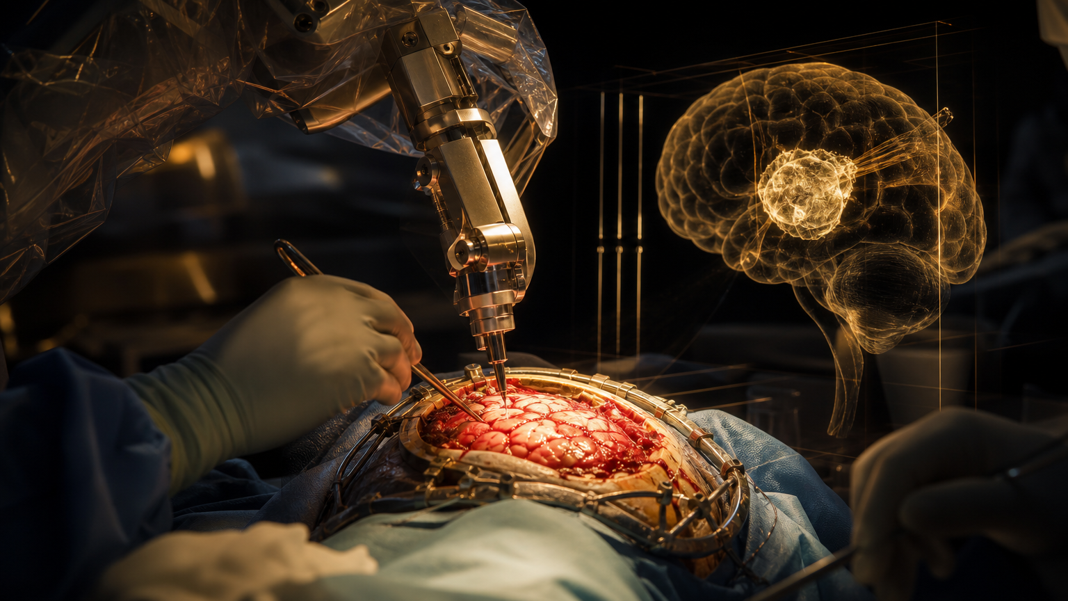
A world-first framed by restraint, not hype
Artificial intelligence has entered one of medicine’s least forgiving environments: brain tumour surgery. Surgeons at the National Hospital for Neurology and Neurosurgery in London, part of University College London Hospitals NHS Foundation Trust, have reported the first live AI-assisted operation to remove a brain tumour, protecting the sight of 48-year-old Rhys Hibbert from Bedfordshire . The operation was carried out as part of a clinical trial using technology developed in-house at University College London and funded by the National Institute for Health and Care Research .
The key point is what the system did, and what it did not do. The AI did not replace a neurosurgeon, make autonomous cuts or take control of the operation. Instead, it analysed the live surgical video feed in real time and highlighted critical structures at the base of the brain, giving the surgical team additional visual information during a procedure in which tiny anatomical differences can change the outcome . UCL described the intervention as the first use of AI to support a neurosurgeon in real time during live surgery on a patient having a brain tumour removed .
The case involved a tumour on Hibbert’s pituitary gland, a small gland at the base of the brain located close to blood vessels and nerves that control vision . UCLH said that, in this area, being a millimetre wrong can make the difference between a successful removal and severe harm, including blindness, stroke or death . That is why the story matters: it is not a general claim that AI can “do surgery,” but a specific example of AI being tested as a precision aid where the margin for error is extremely narrow.
What happened in the operating theatre
Hibbert’s tumour measured about 11 millimetres and had threatened his sight, according to reports from UCLH, UCL and major news outlets covering the announcement . He had been diagnosed after collapsing during a walk and suffering a seizure in 2024, and his symptoms later worsened, including hormone imbalance and vision problems . Without surgery, clinicians said the tumour could have continued to threaten his sight and ultimately led to blindness .
During the procedure, the surgical team used an endoscopic video feed, and the AI system analysed what the camera was showing during the operation rather than relying only on pre-surgical scans . The Guardian reported that the technology helped identify structures such as nerves and blood vessels near the tumour, while the surgical team remained in full control . BBC reporting described the tool as tracking surgical instruments and marking areas where vessels and nerves were likely to be, helping surgeons choose the safest route to remove as much tumour as possible .
Professor Hani Marcus, consultant neurosurgeon at the National Hospital for Neurology and Neurosurgery and professor of neurosurgery at UCL Queen Square Institute of Neurology, performed the first AI-assisted surgery alongside Danyal Khan, the surgical resident leading the work . UCL said the system runs on an NVIDIA Clara IGX platform designed to support real-time AI in medical device settings . The UCL Hawkes Institute developed the AI system, with Dr Sophia Bano, associate professor in robotics and AI at UCL Computer Science, serving as technical lead .
Why this is different from ordinary surgical imaging
Brain surgery already uses sophisticated imaging, navigation and monitoring. The novelty here is the live interpretation of intraoperative video by an AI model trained and evaluated on annotated endoscopic pituitary surgery videos from previous operations . In other words, the system was not simply displaying a scan or pre-planned map; it was helping interpret what the surgeons were seeing in real time .
UCL said the technology had previously been evaluated and used as a training tool for surgeons carrying out this kind of procedure, but this case marked its first live use on a patient . The National reported that the system analysed live surgical video and provided support intended to help the operating team make more precise decisions during delicate work . That shift — from education and research into live intraoperative support — is the substantive milestone.
The system was trained on hundreds of surgical videos, exposing it to a range of anatomy and operative situations that would take an individual surgeon years to encounter, according to UCL’s technical lead . That does not mean the AI is more qualified than a surgeon. It means it can be used as a pattern-recognition layer: a tool that may flag anatomy, instruments and tissue interactions while the human team retains responsibility for interpretation and action .
The patient outcome
The reported patient outcome was striking. UCLH said the operation removed the tumour and protected Hibbert’s vision . Hibbert said his vision had dramatically improved when he woke from surgery, and UCLH reported that within a week he was walking independently without glasses or sticks . The Guardian reported that he has since returned to work as a customer service manager . Global News also reported that the operation restored the sight of the 48-year-old patient and that details were kept private until he had made a full recovery .
For a first case, that recovery narrative will understandably draw attention. But the clinical importance of the case should be measured carefully. A single successful operation is not enough to prove that AI assistance improves outcomes across patients, hospitals and surgeons. The more important next step is the clinical trial process itself: collecting data, testing safety, comparing performance and establishing whether the tool reduces risk in a reproducible way.
The accountability question
The London case lands at a time when health systems are trying to separate useful medical AI from marketing claims. The most reassuring element is that the surgical team remained in control throughout . UCLH and UCL framed the system as decision support, not autonomous intervention . UK Health Innovation Minister James Frith said the case showed AI at its best while also stressing that safeguards and safety must be taken seriously .
That distinction is essential. In an operating room, accountability cannot be blurred. If an AI system highlights an area as risky, the surgeon still has to decide whether the signal is correct, relevant and actionable. If the system misses a structure or produces an uncertain overlay, the surgeon’s anatomical knowledge and judgement remain decisive. The human-in-the-loop model is not a public-relations detail; it is the safety architecture of the procedure.
This also means that adoption cannot be judged by technical performance alone. Hospitals will need evidence about reliability, failure modes, training requirements, integration with existing surgical workflows, cybersecurity, regulatory clearance and informed consent. Patients will need to understand what role AI is playing: not a robot surgeon, not a replacement doctor, but an additional analytical tool used during a high-risk procedure.
A narrow case with broad implications
The most plausible near-term future is not fully autonomous neurosurgery. It is a series of specialised AI systems trained for narrow surgical tasks: identifying structures, warning about danger zones, tracking instruments and comparing the live scene with patterns learned from past procedures. In pituitary surgery, where the operative field is small and critical structures are densely packed, that kind of support could be particularly valuable.
If validated in larger studies, tools like this could help standardise expert techniques and reduce some of the uncertainty that accompanies complex operations. They could also help train younger surgeons by showing how expert-level anatomy recognition plays out during real procedures. But the current evidence remains early: one publicised first patient, one successful operation, and a trial that must now show whether the promise survives broader testing.
The meaning of the London operation is therefore both ambitious and limited. AI has entered brain surgery, but as an assistant. The surgeon still operates, the clinical team still carries responsibility, and the patient outcome still depends on human skill. The breakthrough is that machine vision may now be close enough, fast enough and clinically focused enough to support judgement in real time — precisely where a millimetre can matter.
Sources from the last 72 hours
- [1]First patient in live AI assisted sight-saving brain surgeryAug 27, 2026, 12:00 AM UTC
- [2]First patient in live AI-assisted sight-saving brain surgeryAug 26, 2026, 12:00 AM UTC
- [3]London neurosurgeons perform first successful AI-assisted operation to remove brain tumourAug 26, 2026, 11:01 PM UTC
- [4]UCL surgeons pioneer AI-aided brain tumour operationAug 27, 2026, 9:25 AM UTC
- [5]Surgeons successfully complete 1st-ever AI-assisted brain tumour removalAug 27, 2026, 7:00 PM UTC
- [6]World's first patient to undergo live AI-assisted brain surgery has tumour removedAug 26, 2026, 11:07 PM UTC
AI-generated article based on recent web research, then preserved as a dated editorial snapshot.
Comments
Be the first to comment.